Metaplastic carcinoma (MPC), an uncommon but often aggressive breast cancer, can be challenging to differentiate from other types of breast cancer and even benign lesions based on the imaging appearance. It has a variable pathology classification system.
These types of tumors are generally rapidly growing palpable masses. MPCs on imaging can present with imaging features similar to invasive ductal carcinoma and probably even benign lesions. The purpose of this article is to review MPC of the breast including the pathology subtypes, imaging features, and imaging pathology correlations.
By understanding the clinical picture, pathology, and overlap in imaging characteristics of MPC with invasive ductal carcinoma and probably benign lesions can assist in diagnosing these difficult malignancies. Metaplastic carcinoma (MPC) of the breast is an uncommon form of breast cancer comprising less than 5% of breast cancer patients. It consists of a heterogenous group of malignant neoplasms containing both glandular and non-glandular components with mixed epithelial and mesenchymal differentiation.
The classification of MPCs according to the various systems can be confusing. The World Health Organization classifies MPC into (1) epithelial type and (2) mixed type with further differentiation into subtypes. The more popular Wargotz and Norris classification differentiates MPCs into 5 subtypes: spindle cell, squamous cell, carcinosarcoma, matrix-producing, and MPC with osteoclastic giant cells. The differential diagnosis between invasive ductal carcinoma, MPC, and breast sarcoma is important for management and prognosis. MPC should be differentiated from invasive ductal or invasive lobular carcinoma and primary breast sarcoma in order to determine appropriate treatment options, management considerations, and patient outcome. The pathology classification of MPCs is often variable.
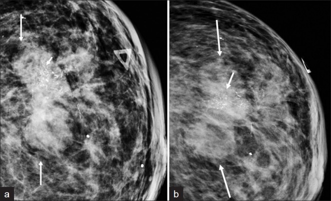
The mammographic, sonographic, and MRI imaging characteristics of MPCs can be similar to invasive ductal carcinoma as well as probably benign features. The imaging appearance reported similar to invasive ductal carcinoma includes an irregular or circumscribed palpable mass with spiculated portion on mammography and solid irregular vs. mixed cystic and solid mass on ultrasound. Prior papers have also reported presentations with imaging features more suggestive of benign masses including circumscribed round or oval masses on mammogram, oval, round, or lobular circumscribed hypoechoic solid mass with posterior acoustic enhancement on ultrasound, and T2 hyperintensity on MRI.
By understanding the clinical picture, pathology, and overlap in imaging characteristics of MPC with invasive ductal carcinoma and probably benign lesions can assist in diagnosing these difficult malignancies.